

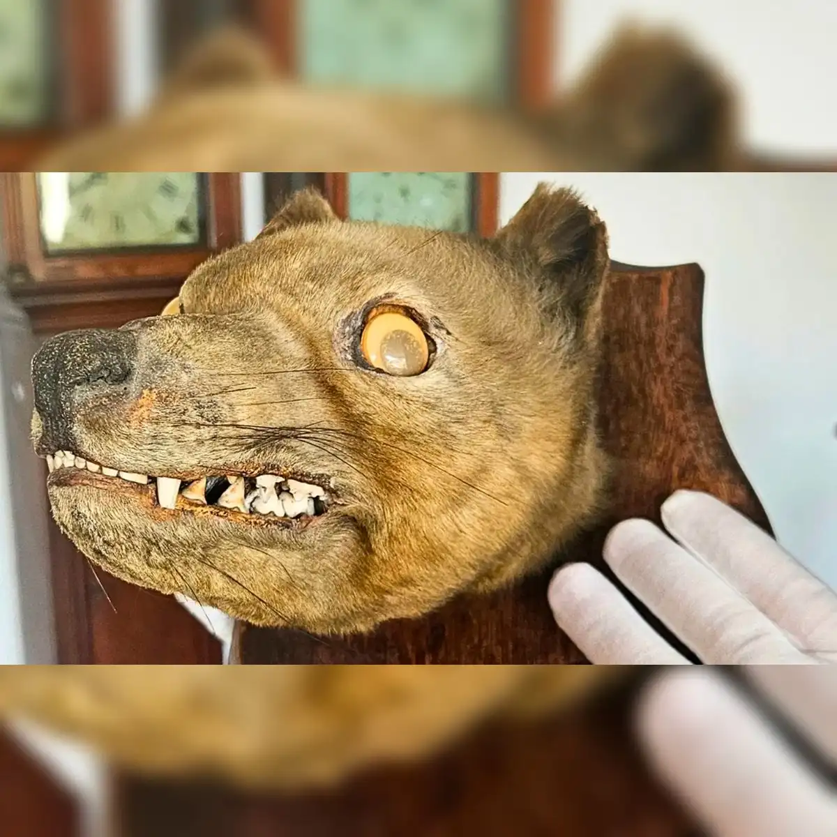
British collector James Cranfield identified a rare mounted head of an extinct Tasmanian tiger after an antiques dealer originally bought the item for 50 pounds, believing it was a 19th-century fox.
The dealer acquired the mounted head at an antiques market as an ordinary piece of Victorian taxidermy. When the item was later listed for sale at an auction, photographs of the lot caught the attention of Cranfield, a British collector who specializes in historical animal mounts.
Cranfield collects unusual fox heads to document the varying techniques and qualities of taxidermy produced by different craftspeople over the decades. However, while examining the enlarged auction photographs, he noticed structural features that did not match those of any known fox or canine species.
The head belonged to a thylacine (Thylacinus cynocephalus), also known as the Tasmanian tiger. The species was a carnivorous marsupial native to Australia, Tasmania, and New Guinea. Decades of intensive hunting, government bounties, and habitat disruption devastated the population, leading to its demise. The last known surviving Tasmanian tiger died in captivity at Hobart Zoo in Tasmania on September 7, 1936.
Dental evidence and auction bidding war
The primary clue that led to the identification was located in the animal's mouth. Members of the canine family, including red foxes, possess six upper incisors. When Cranfield inspected the teeth shown in the high-resolution auction photos, he counted eight upper incisors instead.
The presence of eight upper incisors is a distinct anatomical marker of the thylacine. Although the thylacine shared a similar head shape and jaw alignment with dogs and foxes due to convergent evolution, its dental structure remained that of a marsupial. Marsupials are mammals whose females carry and nurse their young in abdominal pouches, setting them apart from placental mammals.
Cranfield was not the only potential buyer who noticed the anatomical anomaly in the catalog photos. Another collector reached the same conclusion regarding the dentition, setting off a competitive bidding war during the auction.
The rival interest drove the price far beyond its initial purchase price. The auction concluded with a final bid of 62,600 pounds, an amount equal to more than 85,000 US dollars.
Cranfield said that when he clicked to open the larger image, the realization hit his mind and eyes like a lightning bolt. He recognized immediately that the specimen was not a fox and saw that it matched the appearance of a Tasmanian tiger.
Computed tomography and X-ray imaging
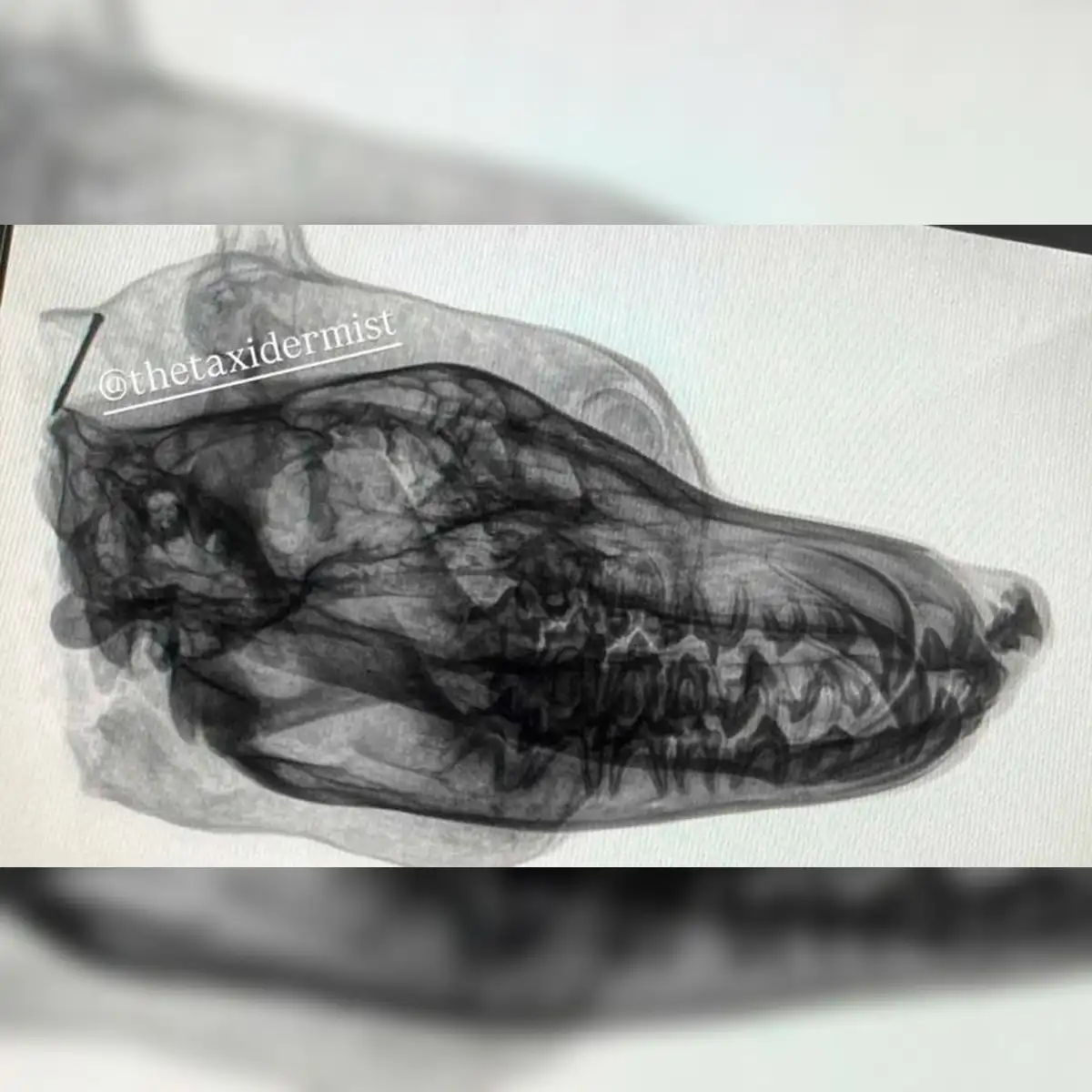
Having committed a substantial sum for the auction lot, Cranfield sought scientific verification of the physical specimen. To confirm that the taxidermist had prepared an authentic animal rather than a composite or altered fox mount, he requested X-rays and computed tomography scans.
Computed tomography, commonly known as CT scanning, uses computer-processed combinations of multiple X-ray measurements taken from different angles to produce cross-sectional images of internal structures. In museum science and paleontology, CT scanning allows researchers to examine delicate internal bones without cutting into historic taxidermy mounts or damaging protective coverings.
The imaging results resolved all doubts by demonstrating that a complete, authentic skull was present inside the mounted head. Experts described the internal skull revealed by the scans as structurally perfect.
The radiographic images provided detailed views of the cranial cavity, nasal bones, and tooth root sockets. The scans showed a noticeably bulging forehead structure characteristic of the species, confirming the physical identification beyond question.
Specialists from the International Thylacine Specimen Database examined the radiographic scans. The organization operates an international scientific repository that catalogs, verifies, and records surviving physical remains of thylacines in public institutions and private hands worldwide.
Stephen Sleightholme, director of the International Thylacine Specimen Database project, told ScienceAlert that he conducted a thorough examination and analysis of the X-ray images, focusing specifically on the skull and tooth arrangement. Sleightholme confirmed that the radiographic evidence offered clear diagnostic support for identifying the specimen as a genuine thylacine.
Scientific significance and genetic research
The discovery carries immense historical and scientific value because it represents only the second taxidermy-mounted thylacine head known to exist globally. Most surviving physical evidence of the extinct species consists of unmounted skulls, isolated bones, or preserved skins rather than finished taxidermy head mounts.
The International Thylacine Specimen Database holds records for more than 800 individual specimens worldwide. The registry includes skeletal remains, wet tissue samples preserved in alcohol solutions, full skins, and microscopic slide preparations. The addition of this newly verified head expands the international inventory of rare physical artifacts.
The specimen offers scientists an opportunity to examine the head shape, cranial contours, and tooth structures of the extinct marsupial in three dimensions. Because the preserved skin covers the underlying bone, researchers can study how soft facial tissues rested over the skull frame.
The taxidermy mount may also allow for genetic testing. Brandon Menzies, a bioscientist at the University of Melbourne, explained that his scientific team could extract approximately 100 milligrams of bone powder from the teeth by performing a minimally invasive drilling technique.
The University of Melbourne is a leading public research institution in Australia with extensive programs in evolutionary biology, genetics, and natural history. Menzies noted that while extracting DNA would not constitute a major breakthrough toward de-extinction, the bone material could broaden scientific understanding of genetic diversity within historical thylacine populations.
Unsolved origins and specimen preservation
It remains unclear how the taxidermy head originally arrived in the United Kingdom. Cranfield suggested that the piece might have been brought to Europe as a hunting trophy or prepared by a taxidermist in Australia before being exported during the 19th or early 20th century.
Researchers believe that future analysis of the timber used for the mounting board could yield critical information. Testing the type and age of the wood backing could help determine where and when the specimen was mounted for display.
Cranfield stated his intention to preserve the outward appearance of the historic mount while making the specimen accessible for scientific research. He aims to support ongoing studies and genetic testing without causing structural damage to the rare artifact.


